Tuesday, May 31, 2016

Perch Dissection

Percidae (Perch) Dissection


Background Information (Jordan)
  • Habitat: Perch live in multiple types of freshwater environments, including warm and cool ponds, lakes, and streams. Perch typically live in shallower waters in these environments, usually never habiting thirty feet below the surface of the water.
  • Diet: Perch feed mostly on invertebrates. When the fish is young, they typically tend to feed exclusively on zooplankton, but as they grow tend to eat larger play, including bloodworm and other large, freshwater invertebrates.
  • Respiratory System: The perch breathes by taking in water through its mouth and gills, and oxygen from the water is transported to blood vessels to be distributed throughout the fish’s body.
  • Fun facts: Female perch lay eggs in long strands that readily stick to underwater plant life or debris from above the water. The eggs are a bright amber in color.

Anatomy (Jordan)
Perch.png


Incision Guide (Jordan)
PerchIncision.png

Resources (Jordan)
Fun facts: http://www.chesapeakebay.net/fieldguide/critter/yellow_perch

Dissection Procedure Video (Jordan and Jason)

Pictures taken and dissection procedure performed by Trejo and Jason

Crayfish Dissection

Astacoidea (Crayfish) Dissection


Background Information (Jordan)
  • Habitat: Crayfish populate a large portion of the planet, living in freshwater lakes and rivers on multiple continents: including temperate regions of Northern and Southern hemispheres, though none are found on Africa or in India.
  • Diet: Crayfish are known for their large, diverse diet. Depending on what aquatic regions they live, they will potentially eat shrimp, freshwater plantlife, small species of fish, worms, insects, plankton, and snails. If kept in captivity, they will eat pretty much anything given to them, so long as it is edible.
  • Respiratory System: Crayfish gills are attached to the legs they walk with, which allows the gills, which are located underneath the crayfish’s tough carapace, to increase the surface area of the gills and allow water to flow in. The oxygen is then transferred into the bloodstream where it can be used for vital body processes.
  • Fun facts: When crayfish molt their carapace, they oftentimes eat it once it has finished shedding. The crayfish do this in order to recover the nutrients that went into growing it in the first place.


Anatomy (Jordan)
Crayfish.png


Incision Guide (Jordan)
CrayfishIncision.png


Resources (Jordan)
Fun Facts: https://sci312crayfish.wikispaces.com/Fun+Facts+about+Crayfish

Dissection Procedure Video (Jordan and Jason)

Pictures taken and dissection procedure performed by Trejo and Jason

Squid Dissection

Teuthida (Squid) Dissection


Background Information (Jordan)
  • Habitat: Typically, most squid reside in marine environments, but some are known to reside in freshwater environments. Some species prefer warmer waters, others colder waters, all at a variety of different depths. The larger the squid, the higher the depths that it resides in. Giant and Colossal squid live very close to the bottom of the ocean. Squid are most commonly found in the North Atlantic regions, as well as offshore California, Hawaii, and the Gulf of Mexico. However, squid have been known to reside in the North Pacific, the Sea of Japan, and the Bering Sea.
  • Diet: Squid often prey on crabs, small fish, squid smaller than themselves, and many varieties of shrimp. Squid capture prey using the suctioning parts of their tentacles, and use their beaks and razor-sharp tongues to grind the prey down into smaller pieces while they are still alive.
  • Respiratory System: The respiratory mechanisms of the squid are tied to its locomotion. Squid move by collecting water in its mantle, and pushing it out to shoot themselves through the water. Squid do not have traditional gills, and instead have gill-like leaflets called lamellae. When the squid pumps water into and out of its mantle, its lamellae become oxygenated and provide the squid with this resource.
  • Fun facts: Squid exhibit sexual dimorphism, but unlike humans female squid at a mature age are larger than their male counterparts. Squid also have large brains in comparison to their body size, making them rather intelligent creatures.


Anatomy (First Picture Trejo, Second Picture Jordan)
Squid.png

Incision Guide (Jordan)
Squidincision.png


Resources (Jordan)
Fun facts: http://www.10-facts-about.com/squids/id/1063

Dissection Procedure Video (Jason)
Pictures taken and dissection procedure performed by Trejo and Jason

Clam Dissection

Veneridae (Clam) Dissection


Background Information (Jordan)
  • Habitat: Most species of clam live in marine environments, and are located in waters of many different temperatures: Florida, Gulf of Mexico, the west coast of North America, around the British islands, and offshore from France. However, many other species of bivalve mollusk—including that of the clam—live in other waters around the world, some even living in freshwater environments; one of which being the Asiatic clam, which occupies many Asian and North and South American rivers, streams, and canals.
  • Diet: Clams feed exclusively using filter feeding. Using their siphons while buried in sediment, the clam is able to capture particles of detritus and plankton, using the mucous-covered surface of the siphon.
  • Respiratory System: Clams breathe through their siphons, respiratory tubes originating from the mantle. Using their foot, they are able to burrow into sand and gravel seabeds, with their siphon protruding from where they burrowed. The siphon then harvests oxygen from the water, as well as capturing food particles using the mucous located on the surface of the siphon.
  • Fun facts: Clams do not have brains, but are programmed to perform only two functions: feeding and reproducing. Clams reproduce by releasing their gametes into the water around them, which forms clam eggs in a process called spawning.


Anatomy (Jordan)
Untitled.png

Incision Guide (Jordan)
ClamIncision.png

Resources (Jordan)
   http://www.asnailsodyssey.com/LEARNABOUT/CLAM/clamFood.php
Dissection Procedure Videos (Jordan and Jason)

Pictures taken and dissection procedure performed by Trejo and Jason

Friday, May 13, 2016

Artificial Selection Lab

Artificial Selection Lab
Purpose:
The purpose of this lab was to attempt to artificially select genes to be expressed in plants. We used a variety of plants expressing different physical traits to be included in our parent generation, then we experimented with various offspring crosses in order to artificially select the traits that the plants would express.

Introduction:
The phenotypes expressed by plants all rely on the genes that the plant carries in its DNA sequence. The genotype (that is, the genes that the plant has in its DNA sequence) is determined by the specific alleles that the parent plants pass down through their gametes to the child organism. For example, if two plants heterozygous for the “tall” gene (which will be considered dominant—T—over the gene for small—t) the child plant will be homozygous tall (TT) one-fourth of the time, heterozygous tall (Tt) one-half of the time, and homozygous short (tt) one-fourth of the time. In this lab, similar questions of heritability were questioned, except concerning multiple phenotypes.

Methods:
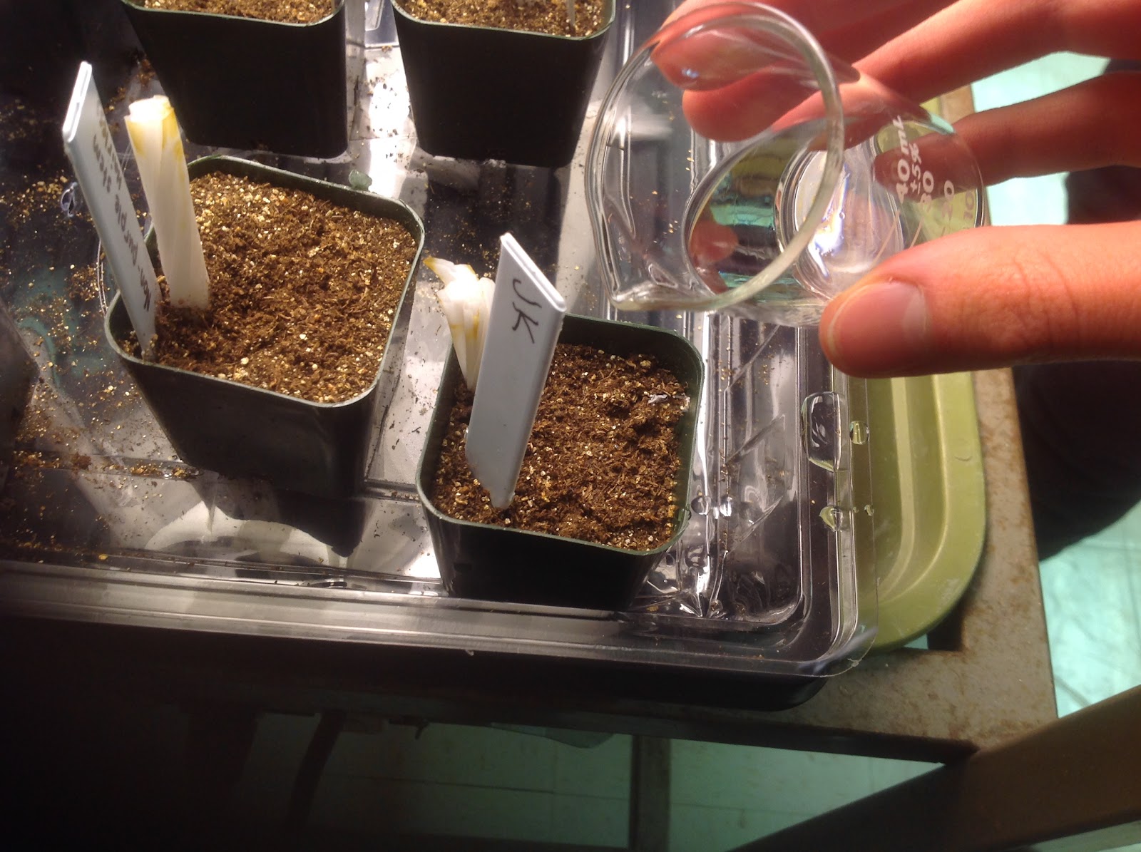
Firstly, we began by setting up the plant pots that would house the seeds that would grow the first generation (F1) of plants. We threaded a wicking cord through a hole in the bottom of the plant pot, filled the pot with light planting mix and packed it in gently. In one pot, we planted six purple stem, hairy seeds, in another we planted six non-purple, hairy seeds, and in a third we planted six non-purple stem, yellow-green leaf seeds.
Fast Plants: The Beginninging

Once that was completed, the seeds were covered in a layer of planting mix, the wicks were threaded into the dilute fertilizer reservoir, and each pot was marked with a label indicating what plants were planted there.
Over the next several days, the plants were watered daily and observed for growth. Here’s what happened over those days...
(The first watering) Fast Plant seeds…

(The first watering) Fast Plant seeds…

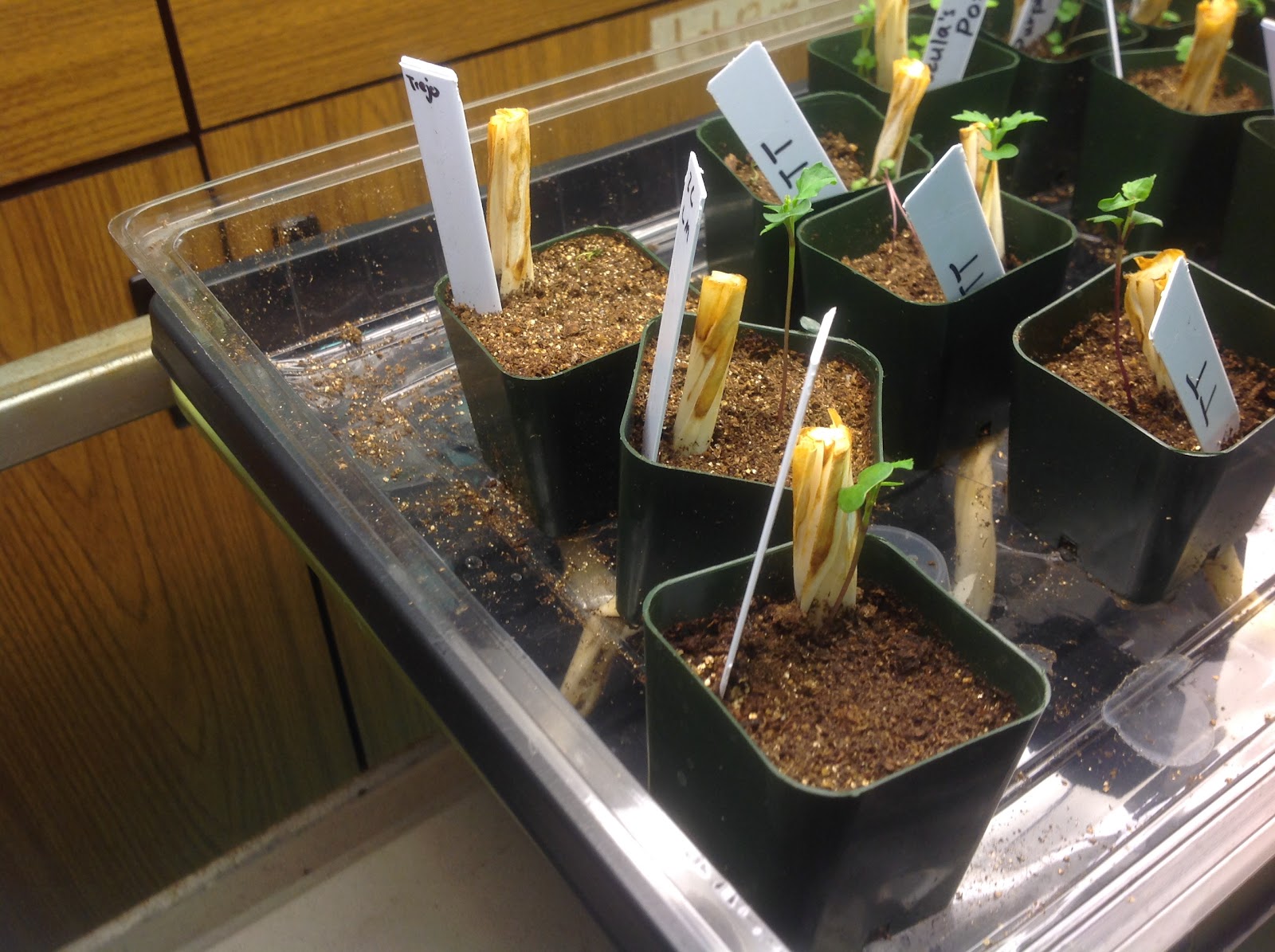
(The first germination) Put ‘em in the soil and watch ‘em grow… HEY!

Sir, we have achieved growth in all species.

However, our victory was short lived. Non-purple stem, yellow-green leaves was the first to die. (Not to mention that only one seed from each seed type grew…)

But yellow-green soon made an attempt to rebound…

Only to fail once again. RIP in pastas our dear friend yellow-green.

Filled with determination, the hairy and the non-hairy continued to grow.

Only for the worst to happen… Hairless joined the yellow-green family in death. Buuuut on the bright side our purple-stemmed fellow blossomed!

Only the strong survive.

With the memory of our fallen kin fresh in the back of our minds, we had no choice but to continue the experiment with the sole remaining plant our group had… the purple-stemmed, hairy plant. However, in order for us to attempt cross pollination between different plant species to determine the phenotypes of the F2 generation, an outside plant would need to be used to produce seed pods.
As such, we used a living, non-purple, yellow-green leaf plant from another group to facilitate breeding. Using a cotton swab, we gathered pollen from the yellow-green leaved plant and deposited it onto the flowers of our purple-stemmed, hairy plant. After that was done, we waited for seed pods to grow.
Gathering plant sperm has never been more fun.

The soil labels from both plants.

Swab o’ sperm.

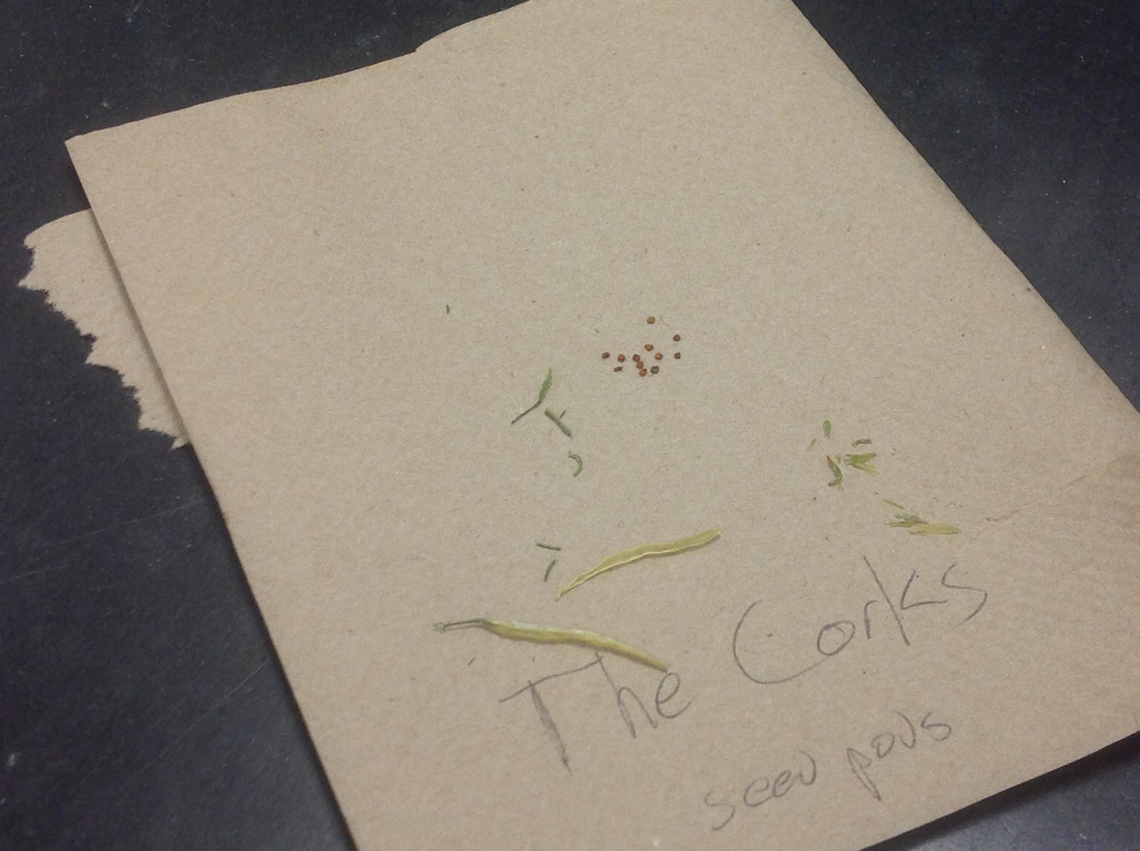
After several more days, seed pods were had! If our hypothesis was right, these seeds would have the potential to grow both yellow-leaved, non-purple stemmed plants and purple-stemmed, hairy plants. Depending on how the genes are organized on the chromosome, there would potentially be hairy, non purple-stemmed plants, yellow-green, purple stemmed plants, etc. After removing the seed pods from the plant, we euthanized the plant and allowed the seed pods time to dry out.
The fruits of our labor.

The seeds after a weekend of drying out.

RIP our sole survivor.

Once we gave the seeds time to dry, we repeated the earlier steps of preparing a plant pot, and planted six of the seeds from our seed pods in the prepared pot. After that, we gave our F2 generation time to grow and develop.
Preparing a new plant pot for a new generation of survivors!

Unfortunately, however, the seeds never grew, and we had no F2 generation to analyze. And thus, the reign of the purple, hairy-stemmed plant came to an end.
A graveyard of seeds.

Data:
Because the F2 generation of plants wasn’t able to germinate, there isn’t exactly much data to be gleaned from this lab. We cannot determine how the phenotypes relate to their position on the gene because we could not determine which plants would grow from a Purple Stemmed, Hairy x Non-Purple Stem, Yellow-Green Leaf testcross. Even within the first generation, the fact that only one plant grew curtails any comparison that could be made between F1 plants. As such, we can only really hypothesize as to what plants the aforementioned testcross would be able to produce. If each of the characteristics of the plants follows simple Mendelian dominance, whichever is dominant in height, leaf color, hairiness or lack thereof, and stem color would occur in a 3:1 ratio favoring the dominant trait, provided that the dominant traits were heterozygous. If all the dominant traits were homozygous, the F2 generation would show only show dominant traits. If the genes for specific traits were linked, however, most of the plants would look like the F1 generation, but a few would have characteristics of both parent plants. The offspring would appear in a 9:3:3:1 ratio, favoring plants that appear most similar to the F1 generation.

Discussion:
Altering the environmental conditions would affect the rate of survival for plants with certain characters. In our experiment the purple stem hairy plant was better equipped to survive in these conditions. For example, in harsh conditions hardier plants will survive. With artificial selection one can eliminate a trait. If we chose to remove the hairy characteristic of the plants we are able to do this by breeding the two non hairy plants and leaving the purple hairy stem out of the cross for several generation until we don't see that trait anymore.

Conclusion:
In the first generation of plants we planted three seeds of all three types of plants: purple stem hairy seeds, non-purple stem yellow green leaf, and non-purple hairless seeds. At first we started with dirt, then we planted the offspring of the F1 generation. For the F2 generation we planted 6 seeds and unfortunately none grew. We predicted that the reason why the F2 generation couldn't reproduce was because the lack of water, too much water, not enough light from the UV, too much light from the UV, mot enough nutrients in the soil, and competition between the 6 seeds. We also believe that the seed pots that were placed at the corner of the tray were at a disadvantage because they didn't get as much light to grow. However, our F2 generation of plants were in the middle of the tray, so whether or not that is an accurate hypothesis is indeterminate.

References:

  • Unless otherwise stated, all information related to artificial selection has been synthesized from Jane Reece’s Campbell Biology AP Edition Textbook and the BioFax! Artifical Selection Lab from Flinn Scientific Inc.
  • Paraphrased procedures as shown in the Methods section have been synthesized from procedures found in BioFax! Artifical Selection Lab from Flinn Scientific Inc.
  • All pictures are original from the in-class experiment.

Sunday, April 10, 2016

AP Bio Peppered Moth Quest

Jason Lopez


Peppered Moths


Evolutionary changes in population are almost always directly related to changes in the environment. In the case of Peppered Moths, a large scale shift in External coloring was observed. This shift was characterized by a rapid depletion of light colored moths and increasing numbers of Dark Colored Moths reaching almost 98% dark colored. Coincidentally, this shift was observed during the English Industrial Revolution, a time in which Coal power was heavily utilized, leaving the English Forests blackened with Soot. Because Dark Moths are better suited to survive in the newly blacked forests due to lessened visibility to predators, their numbers began to outrank those of the Light colored Moths. This change in Moth color can be directly attributed to Human meddling in the Ecosystem, leaving soot across the countryside, discoloring the Country's forests resulting in a lower mortality rate for darker colored Moths. In turn, this lower mortality rate was met by a higher mortality rate for Light colored moths in these sooty forests, resulting in a rapid boom in Dark colored moths.

Saturday, March 19, 2016

Gel Electrophoresis Lab

Jason
Trejo
Jordan

Purpose: The purpose of this lab was to run DNA through the process of gel electrophoresis using 3 restriction enzymes in order to characterize DNA. We were then meant to prepare and analyze our results based on the data we procured by running the DNA through the gel


Introduction: Restriction mapping is used to determine the size of DNA fragments. In gel electrophoresis, the smallest fragments will always travel a larger distance and the bigger fragments will travel a shorter distance.  The negatively charged end of the gel electrophorese repels the DNA moving it towards the positive end. Restriction maps of DNA are the equivalent to fingerprints, put alongside Lambda DNA as a size comparison and control, we were able to characterize the alien DNA.

Methods:
First of all we obtained a gel from our teacher and then we delicately proceeded to load the tiny wells with the contents of each reaction tube, containing DNA.  
(The prepared enzyme/DNA solutions and the Agarose gel.)

With my super technique I was able to skillfully use a needlepoint pipet to load the wells of the gel, being careful that the pipet is void of any air bubbles that could spill the solution.
(Agarose gel after DNA has been administered.)
(Agarose gel being cast into the electrophoresis tank)
The negatively charged phosphate backbone of the DNA allows for it to move through the pores of the heated Agarose gel, towards the positively charged anode at the opposite end of the wells. The lighter fragments move farther because they can squeeze through pores more easily and are faster due to their lighter weight.
   
The completed gel electrophoresis under an orange lamp that allows us to have a better look at where the fragments ended up on the gel. We used sharpie on a plastic Baggie to emphasize the cut points and label the respective enzyme/DNA solutions for easier reference. Cut sites had to be estimated, so the numbers were not adding up to be equal, however the concept and relative lengths of each fragment was understood and noted.

Data:


Discussion: Through gel electrophoresis we were able to obtain enough data to map the plasmid and discovered that the DNA sequence was cut by the restriction enzymes on several occasions. There were two PstI sites , one SspI site, and one HpaI site on the plasmid. Compared to the Lambda DNA, this mystery DNA had much shorter fragments when PstI was administered.


Conclusion: Although our cut sites and fragment lengths did not necessarily add up correctly, we were still able to insert estimated numbers and create a plasmid displaying the data we procured. We were able to determine the enzyme cut sites alone, and in relation to each other during the double and triple digests. The smaller bands move through the gel more quickly due to their size, so we were able to make educated guesses on the positions of cut sites with given number data.